| HOME About Dr. Janson APPOINTMENT FORM Directions Health History Form Links Online Payments Patients' Comments Dental Advice Index |
|
|
|
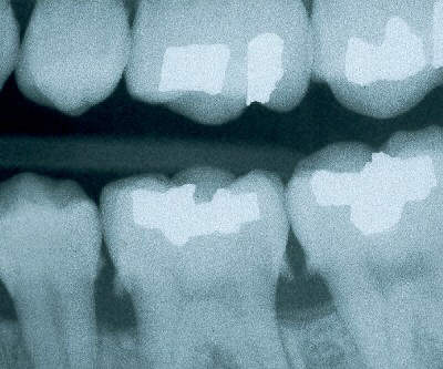
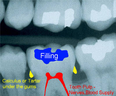
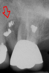
The x-ray above represents a patient who has not visited the dentist for 2 years. The calculus or tartar is now visible on the x-ray as wisps or thorns projecting from the sides of the teeth UNDERNEATH THE GUMS where you can not see them. These 'thorns-of-calculus' are a major cause of puffy and bleeding gums ( gingivitis ) , which leads to bone loss and periodontal disease. Please visit our page on Ultrasonic Cleanings for before and after photos to see how we can make your teeth brighter and squeaky clean once again!
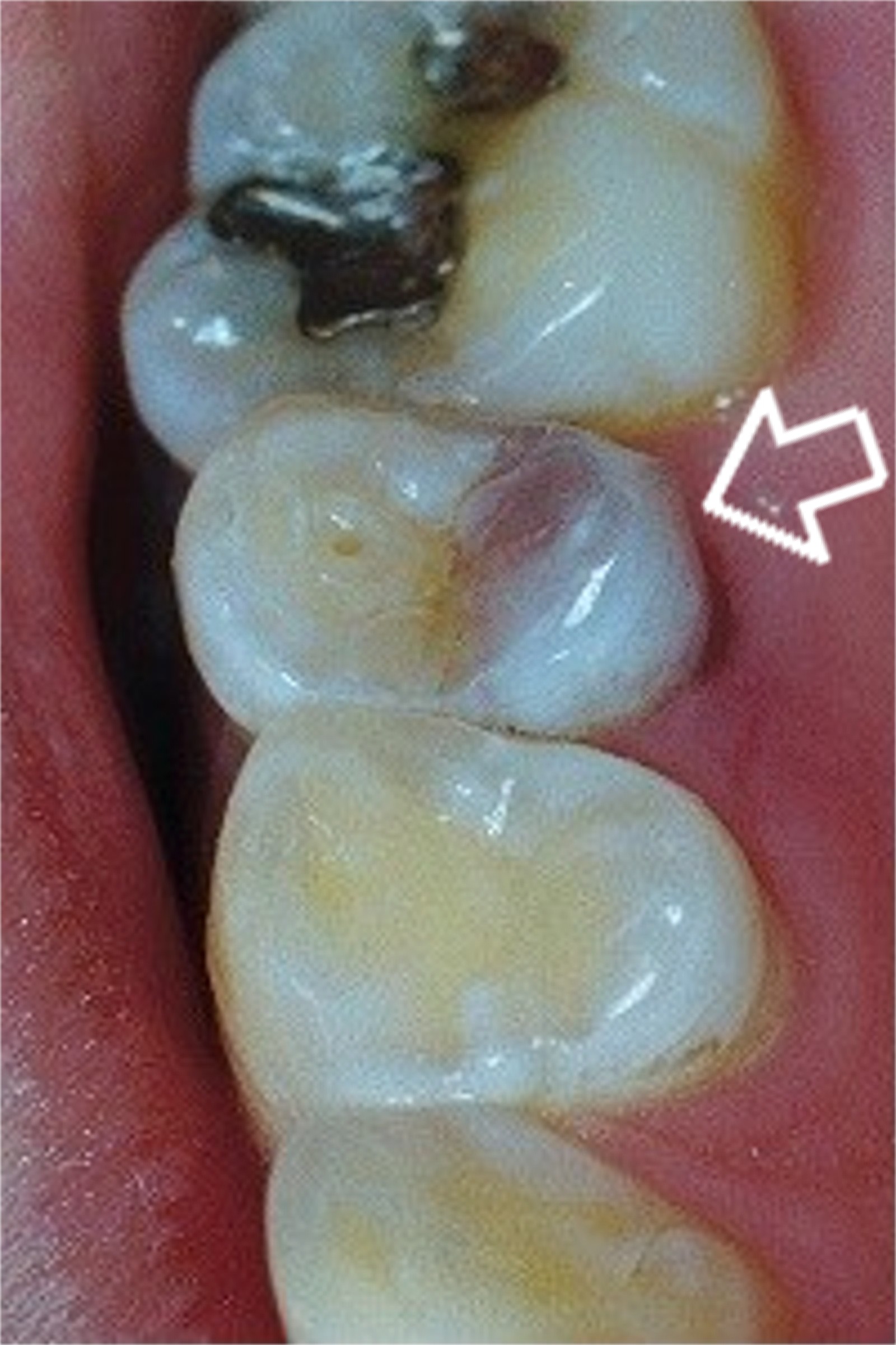
Necrotic Tooth - a tooth which has died
Though many teeth which die do NOT discolor, the dark purple area of this premolar indicates that the pulp of the tooth has died, likely due to a cavity or other trauma. Such a tooth needs root canal or other treatment in a timely manner to prevent extensive infection and abscess. Once a tooth has died, it tends to become brittle and will eventually break down without proper treatment from your dentist. Since the tooth has died, it may not feel sensitive to the patient and is often ignored. As a dentist, I routinely see patients who have broken off the entire crown of a necrotic tooth! Though the tooth may still be saved (ie- see our Cosmetic Dentistry page) after it has broken off, it may also need to be extracted.
|
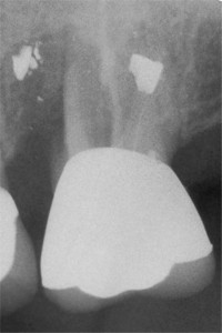
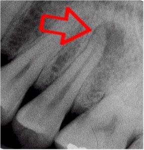 The dark shadow around the root of this tooth indicates a tooth abscess or infection |
Tooth Root Amputation - Without Extracting the tooth!
If the proper conditions exist, we can section and extract the problematic root of a tooth, keeping the rest of the tooth intact! This patient fractured one of the 3 roots of an upper molar and elected to have the problem root removed. The tooth was then smoothed and a filling placed. The white spots at the ends of the roots represent fillings from a prior procedure called an 'apical retrofill'.
I look forward to addressing your immediate and future dental needs and answering any questions you might have.
Ethan Janson DDS
![]()
Dental Advice Index ![]() | Cosmetic Cases / Photos | Our Philosophy
| About Dr. Janson | Appointment Request | Directions | Health Form Printout | Patient Comments | Dentist and Seattle links | Recommend us to a friend | Ask Dr. Janson a question! | Online Payments
| Cosmetic Cases / Photos | Our Philosophy
| About Dr. Janson | Appointment Request | Directions | Health Form Printout | Patient Comments | Dentist and Seattle links | Recommend us to a friend | Ask Dr. Janson a question! | Online Payments ![]()
| Back to Top |
This web site designed by...
![]()
City Dental
Referral Service & Web Site
Design - Dentists Click Here!
©1999-2005 CityDRS.com (City Dental Referral Service and Web
Design)
Last revised: January 09, 2006.